
Large Deep Abscess on the Pastern
ABSTRACT
Transdermal pharmaceutical carbon dioxide was used as an adjunct therapy for a very deep wound on the right rear pastern. Monkey had a severe sub-solar abscess that erupted above the coronary band on the caudo-lateral aspect of the pastern. It was very deep, necrotic and exposed the lateral collateral cartilage. Pharmaceutical carbon dioxide significantly shortened the healing time.
Clinician: Patricia Blakeslee, VMD
Client Information: Flying Monkey - 1995 Thoroughbred gelding
CASE HISTORY AND CLINICAL SIGNS
Signs: On January 16, it was noticed that Monkey was lame right hind (RH) with some pastern swelling. As there was heat in the hoof, the owners presumed that he had a subsolar abscess and proceeded to soak the hoof in warm Epsom Salts. On January 22, the abscess started to erupt about 3cm above the lateral heel. The defect continued to enlarge and was odiferous and necrotic. On presentation on January 23, Monkey was 4-5/5 degrees lame and there was a large area of necrotic tissue with a very foul odor. His temperature was 98.6° F and the physical exam was otherwise not remarkable. I debrided the necrotic tissue; flushed a deep cavity; packed it with Cephaperin Benzathine (Tomorrow treatment for dry cows) and bandaged with Silver Sulfadiazine (SSD). He was placed on Ceftiofur (Excede), Gentamicin, Flunixin meglumine (.5 mg/kg BID) and Naquasone. The owners were instructed to change the bandage daily.
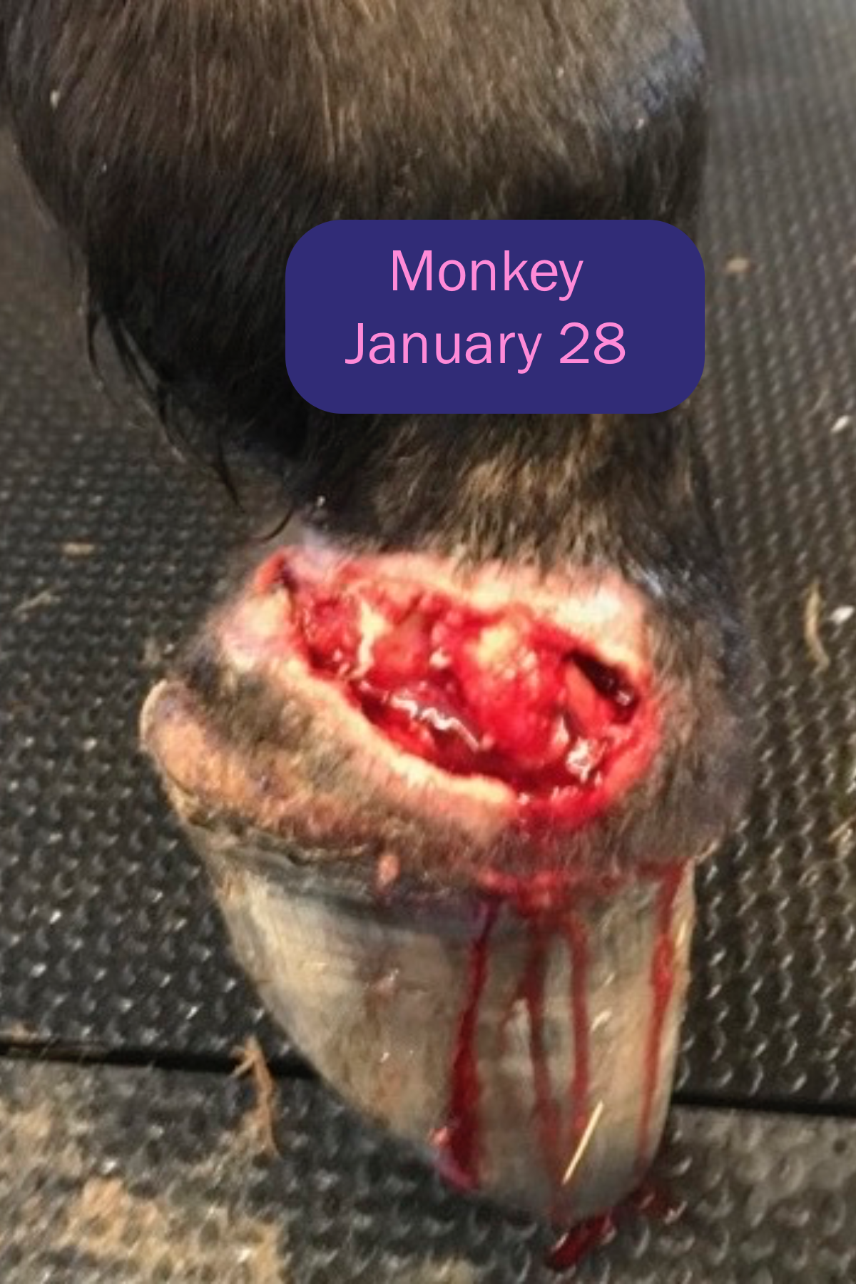
On January 28, he was walking a little better but the large wound gaped every time he tried to put the heels down. There was an additional eruption at the coronary band of the lateral heel. I debrided the remainder of the necrotic tissue. The tip of the lateral collateral cartilage was exposed and the wound extended very deeply into the heel towards the navicular bone. A healthy granulation tissue bed was forming nicely. Radiographs were not remarkable. I shortened the toe to ease breakover so the wound would not gape so much. The flunixin was discontinued and 57mg Firocoxib (Equioxx) a day was started.
Transdermal carbon dioxide was started on January 30th and administered daily for 5 days. The protocol going forward was every other day for 3 treatments and then every third day for three weeks.
On February 13, he was walking well. The crevice was 1.5cm deep and just the tip of the collateral cartilage was exposed. It was mildly odiferous as the bandages were constantly wet due to extensive rain and mud and we were unable to keep him on stall rest due to his personality.
On February 22, the discharge was much less, the overall dimensions were noticeably smaller and he would stand on the RH and rest the LH. The crevice was 3mm deep and even though the collateral cartilage was covered over, the granulation tissue was tenuous.
On March 1, Monkey was very comfortable moving around the pasture in all gaits. The crevice was minimal. I trimmed some exuberant granulation tissue that had developed. All meds were discontinued at this point though bandaging was continued to deter further exuberant granulation tissue.


CONCLUSION
Pastern wounds typically take a very long time to heal and this wound which was not only large, also extended deeply in to the soft tissues. The exposure of the collateral cartilage was also a complication that could have slowed healing or required surgery if it became infected. This is fully healed in 5 weeks. I feel that 5 weeks from abscess eruption to coverage of the collateral cartilage was very quick and was definitely aided by the transdermal carbon dioxide.